


Understanding PET Scan, Biopsy, and Imaging Tests in Cancer Diagnosis
Hearing the word “cancer” often brings a flood of questions.
Many people expect a diagnosis to come from a single test. In reality, cancer diagnosis usually involves several steps. Doctors often combine scans, imaging studies, laboratory findings, and tissue testing before reaching a clear conclusion.
This process can feel confusing, especially when terms like PET scan, biopsy, CT scan, MRI, or screening tests begin appearing during consultations.
Understanding what these tests do can make the journey less overwhelming.
Each one serves a different purpose. Some help detect abnormalities. Others show where a disease is located. Certain tests help determine how far a condition has spread. A biopsy, meanwhile, often provides the final confirmation.
Together, these tools help doctors make informed decisions about diagnosis and treatment.
Why Multiple Tests Are Often Needed
Cancer does not behave the same way in every person.
Some cancers produce symptoms early. Others remain silent for months or even years. Certain growths appear clearly on scans, while others require closer examination before a diagnosis becomes certain.
Because of this, doctors rarely rely on a single investigation.
A scan may reveal an abnormal area. Another imaging test may provide more detail. A biopsy may then be performed to determine exactly what type of cells are present.
Each step helps answer a different question.
The goal is not only to find a cancer if it exists, but also to understand its location, size, and characteristics.
What Imaging Tests Do
Imaging tests help doctors look inside the body without surgery.
Different scans provide different kinds of information.
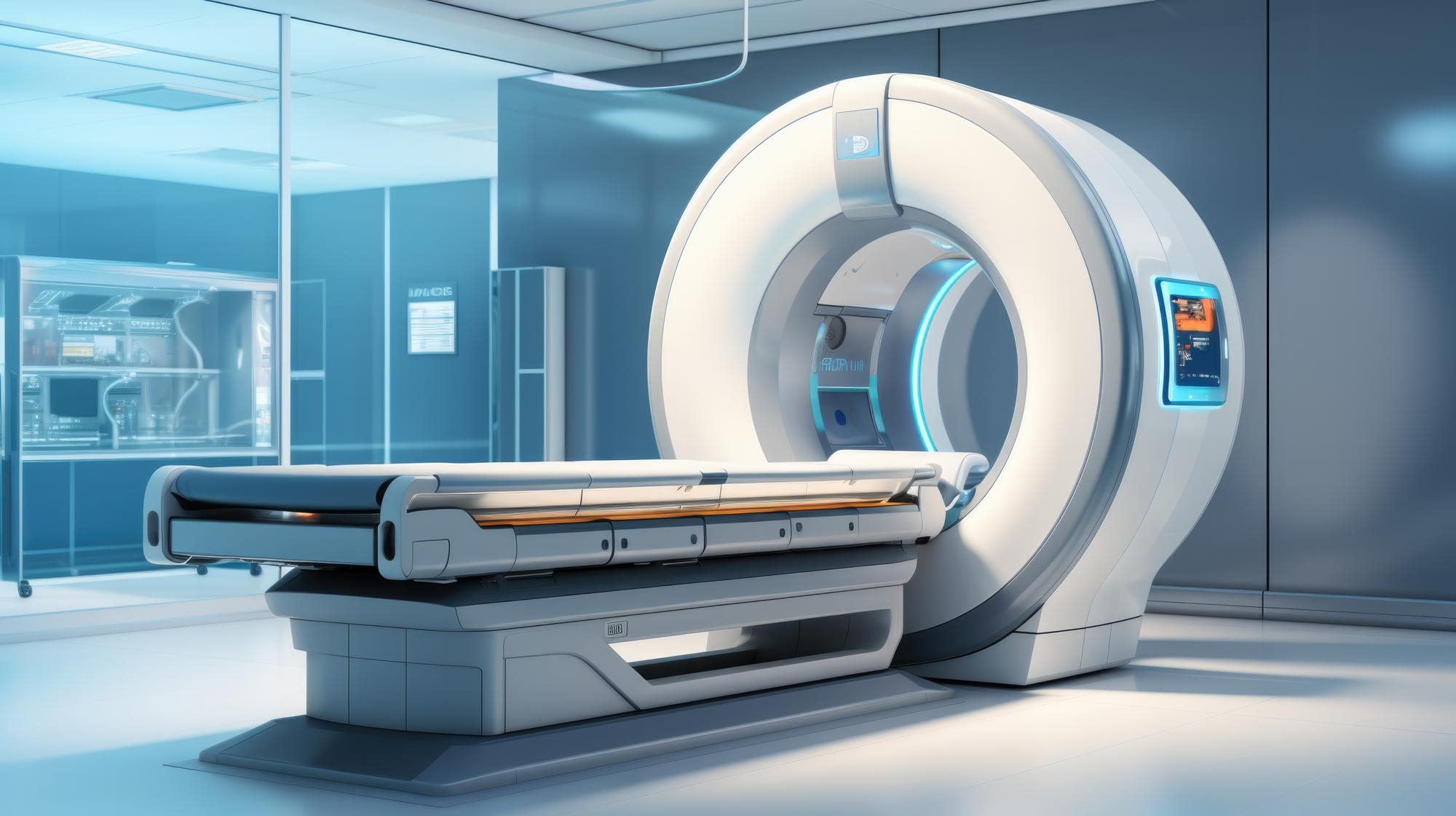
A CT scan creates detailed cross-sectional images of internal organs and tissues. It can help identify masses, enlarged lymph nodes, or suspicious areas that may require further evaluation.
MRI scans use magnetic fields to produce highly detailed images, especially useful for certain soft tissues and organs.
Ultrasound examinations are often used to evaluate specific organs and guide further investigations when needed.
These tests do not always confirm cancer.
What they often do is identify areas that deserve closer attention.
That information helps determine the next steps.
What Is A PET Scan?
PET stands for Positron Emission Tomography.
A PET scan works differently from many other imaging tests because it focuses on how tissues are functioning rather than simply how they look.
During the scan, a small amount of radioactive tracer is introduced into the body. Areas with unusually high metabolic activity may absorb more of the tracer and appear more prominently on the images.
Since many cancer cells grow and divide rapidly, they often show increased activity on PET scans.
Doctors may use PET scans to:
-
Identify suspicious areas
-
Evaluate whether cancer has spread
-
Assess response to treatment
-
Help plan future treatment strategies
Not every abnormal PET scan indicates cancer.
Inflammation and certain infections can sometimes produce similar findings.
That is why PET scans are usually interpreted alongside other tests.
Why A Biopsy Is Often The Most Important Step
Scans can raise suspicion.
A biopsy helps provide certainty.
During a biopsy, a small sample of tissue is removed and examined under a microscope by a pathologist.
This allows doctors to see exactly what type of cells are present.
The results can determine:
-
Whether cancer exists
-
The specific cancer type
-
How aggressive it may appear
-
Additional characteristics that may influence treatment
For many cancers, a biopsy remains the definitive step in diagnosis.
Without tissue confirmation, imaging findings alone may not provide all the answers.
The Role Of Cancer Screening
One of the most important advances in modern medicine is screening.
Screening aims to detect abnormalities before symptoms appear.
In some cases, cancer can be identified at a much earlier stage through routine evaluation rather than after symptoms develop.
Understanding when to undergo cancer screening tests often depends on factors such as age, family history, medical conditions, and individual risk factors.
Regular screening may help identify changes before they become more difficult to treat.
The timing of screening is not the same for everyone.
Personalized medical advice plays an important role.
Screening For Common Cancers
Different cancers require different screening approaches.
For example, colon and rectal cancer screening and diagnosis often involves tests designed to identify abnormal growths or early-stage disease before symptoms develop.
Similarly, mammography remains a key tool in early breast cancer detection and care, helping identify changes that may not yet be noticeable through symptoms alone.
Certain individuals may also require evaluation for upper digestive tract cancers depending on their symptoms, family history, or risk factors. Discussions around stomach cancer screening and treatment options often become relevant when persistent digestive symptoms or specific risk factors are present.
The purpose of screening is simple.
Finding problems earlier generally creates more opportunities for successful treatment.
Why Early Diagnosis Matters
Cancer treatment has advanced significantly over the years.
Many cancers are now highly treatable, particularly when detected early.
The challenge is that early-stage cancer often causes few symptoms.
That is why imaging tests, screening programs, and biopsies play such an important role.
They help identify conditions that may otherwise remain hidden.
Earlier diagnosis frequently allows treatment to begin before the disease progresses further.
Conclusion
Cancer diagnosis rarely depends on a single test. Instead, doctors often combine imaging studies, PET scans, biopsies, and screening evaluations to build a complete picture. Each investigation contributes different information, helping determine whether cancer is present and guiding future treatment decisions.
At KKR Hospital, understanding the purpose of these tests helps patients approach the diagnostic process with greater confidence. While terms like PET scan, biopsy, CT scan, or MRI may sound intimidating at first, each plays an important role in helping doctors identify disease accurately and begin the most appropriate care as early as possible.